![]() Figure 3 of
Wistow, Mol Vis 2002;
8:205-220.
Figure 3 of
Wistow, Mol Vis 2002;
8:205-220.
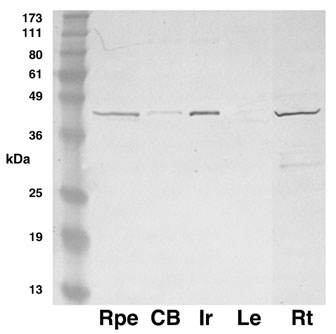
Figure 3. Western blot of Ocsp
Extracts of human eye tissue solubilized in SDS, separated by SDS-PAGE, blotted and hybridized with antiserum Oscp1. Marker sizes are shown. Lanes are Rpe/choroid (Rpe), ciliary body (CB), iris (Ir), lens (Le) and retina (Rt). Extraneous lanes between Le and Rt were cropped from the image without showing white space.