![]() Figure 6 of
Wistow, Mol Vis 2002;
8:171-184.
Figure 6 of
Wistow, Mol Vis 2002;
8:171-184.
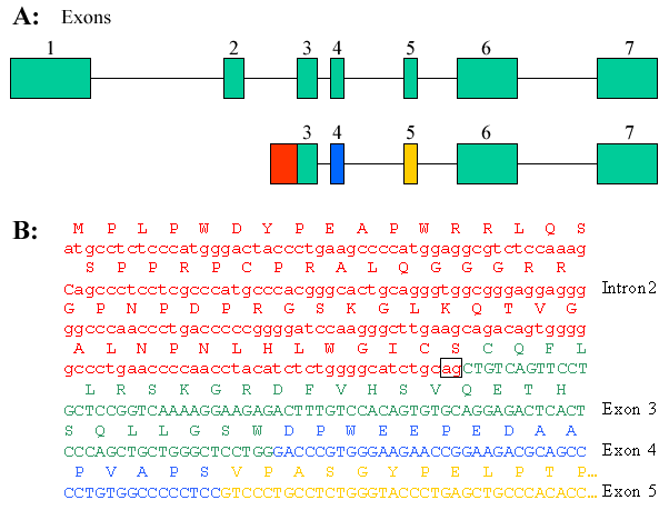
Figure 6. Livin splice variant
A: The structure of the Livin gene on chromosome 20q13.33 is shown with the seven exons numbered. The gene structure corresponding to clone by15h01, which includes sequence from intron 2 is shown in red. B: Partial sequence of clone by15h01, showing ORF running in frame from intron 2 sequence into exon 3, followed by normal splicing to exons 4 through 7. The splice junction for the 5' end of exon 3 in canonical Livin is boxed. Exons are colored in A and B for clarity.