![]() Figure 9 of
Kinkl, Mol Vis 2002;
8:149-160.
Figure 9 of
Kinkl, Mol Vis 2002;
8:149-160.
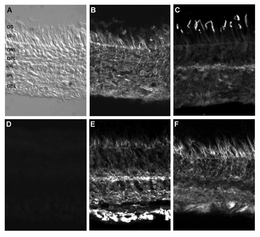
Figure 9. Distribution of FGFR in human retina ex vivo
Human retinal sections taken from the central posterior field, within 5 mm of the optic nerve head (shown by differential interference contrast microscopy in A) were stained with antibodies specific for FGFR-1 (B), FGFR-2 (C), FGFR-3 (E), and FGFR-4 (F). D: Representative control section in which the primary antibody (in this case FGFR-2) was pre-adsorbed with the corresponding immunizing peptide prior to labeling. Explanations of labeling patterns are given in the text. Abbreviations used: GCL, ganglion cell layer; INL, inner nuclear layer; IPL, inner plexiform layer; IS, photoreceptor inner segments; ONL, outer nuclear layer; OPL, outer plexiform layer; OS, photoreceptor outer segments. Magnification x200.