![]() Figure 5 of
Kinkl, Mol Vis 2002;
8:149-160.
Figure 5 of
Kinkl, Mol Vis 2002;
8:149-160.
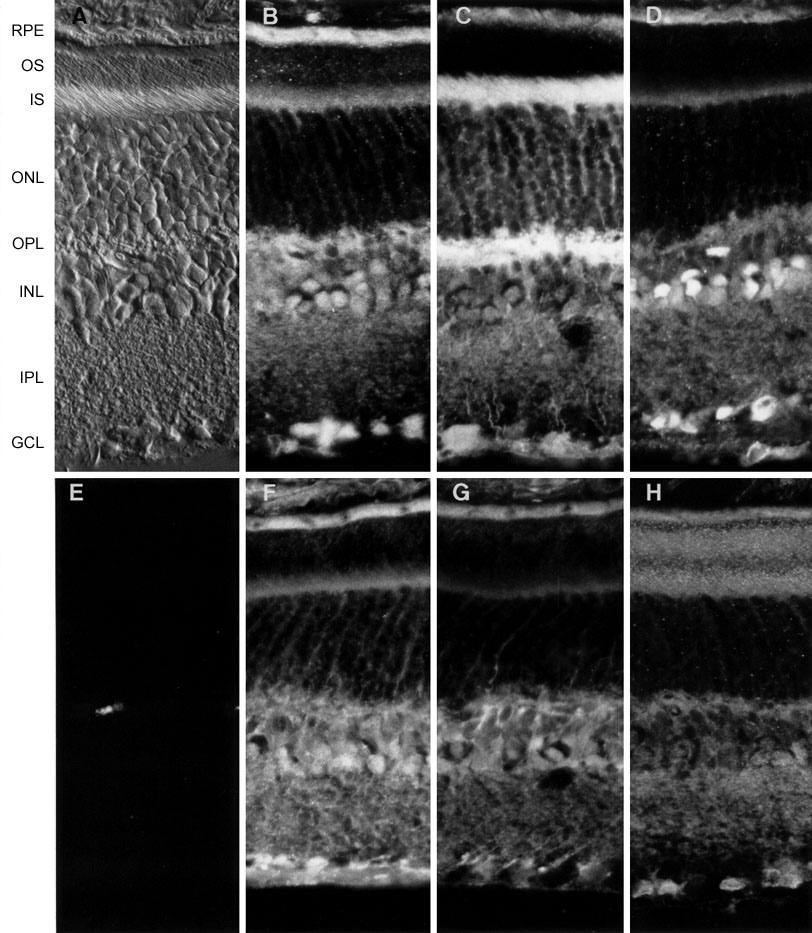
Figure 5. Distribution of potential FGF-related signaling molecules in adult rat retina ex vivo
Retinal sections taken close to the optic nerve head (shown by differential interference contrast microscopy in A) were immunolabeled with antibodies specific for PLCg1 (B), SOS1 (C), SOS2 (D), ERK1/2 (F), SH-PTP2 (G), and Shc (H). Detailed comments on staining patterns are given in the text, but note the difference in SOS1 and SOS1 immunoreactivity. E: Representative control section in which the primary antibody (in this case PLCg1) was pre-adsorbed with the corresponding immunizing peptide prior to labeling. Abbreviations used: GCL, ganglion cell layer; INL, inner nuclear layer; IPL, inner plexiform layer; IS, photoreceptor inner segments; ONL, outer nuclear layer; OPL, outer plexiform layer; OS, photoreceptor outer segments; RPE, retinal pigmented epithelium. Magnification x280.