![]() Figure 4 of
Kinkl, Mol Vis 2002;
8:149-160.
Figure 4 of
Kinkl, Mol Vis 2002;
8:149-160.
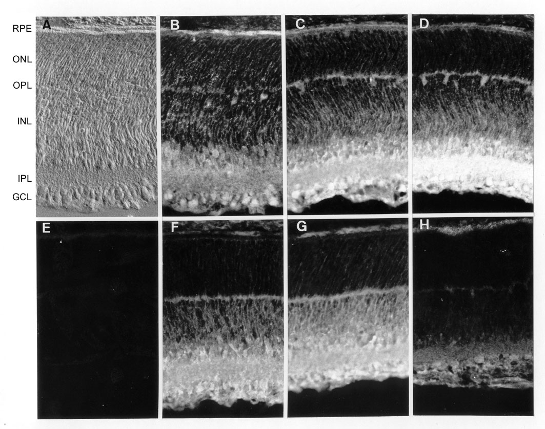
Figure 4. Distribution of potential FGF-related signaling molecules in PN5 rat retina ex vivo
Retinal sections taken close to the optic nerve head (shown by differential interference contrast microscopy in A) were immunolabeled with antibodies specific for PLCg1 (B), SOS1 (C), SOS2 (D), ERK1/2 (F), SH-PTP2 (G), and Shc (H). PLCg1 showed a uniform expression throughout the retina. SOS1 and SOS2 were predominantly localized in the inner retina, fainter labeling being observed in the outer plexiform layer (OPL) and horizontal cell bodies. ERK1/2 and SH-PTP2 were similar in expression, intensely labeling the inner retina. Expression was also found in the OPL and more weakly in the outer nuclear layer (ONL). Only faint Shc labeling was observed, mostly within cells of the ganglion cell layer (GCL) and on the newly forming photoreceptor outer segments. Fainter labeling was observed in the inner plexiform layer (IPL). E: Representative control section in which the primary antibody (in this case SOS1) was pre-adsorbed with the corresponding immunizing peptide prior to labeling. Additional abbreviations: INL, inner nuclear layer; RPE, retinal pigmented epithelium. Magnification x280.