![]() Figure 3 of
Kinkl, Mol Vis 2002;
8:149-160.
Figure 3 of
Kinkl, Mol Vis 2002;
8:149-160.
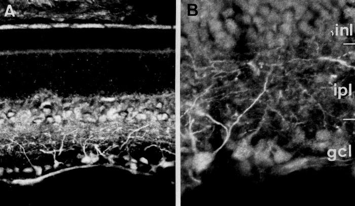
Figure 3. FGFR-3 distribution in adult rat retina ex vivo
Retinal sections were stained with an antibody specific for FGFR-3. A: Lower magnification (x350) image showing predominant expression in the inner nuclear layer (INL), inner plexiform layer (IPL), ganglion cell layer (GCL), and nerve fiber layer (NFL). B: Detail of the inner retina, showing particularly prominent staining of dendrites. Magnification x1000.