![]() Figure 2 of
Kinkl, Mol Vis 2002;
8:149-160.
Figure 2 of
Kinkl, Mol Vis 2002;
8:149-160.
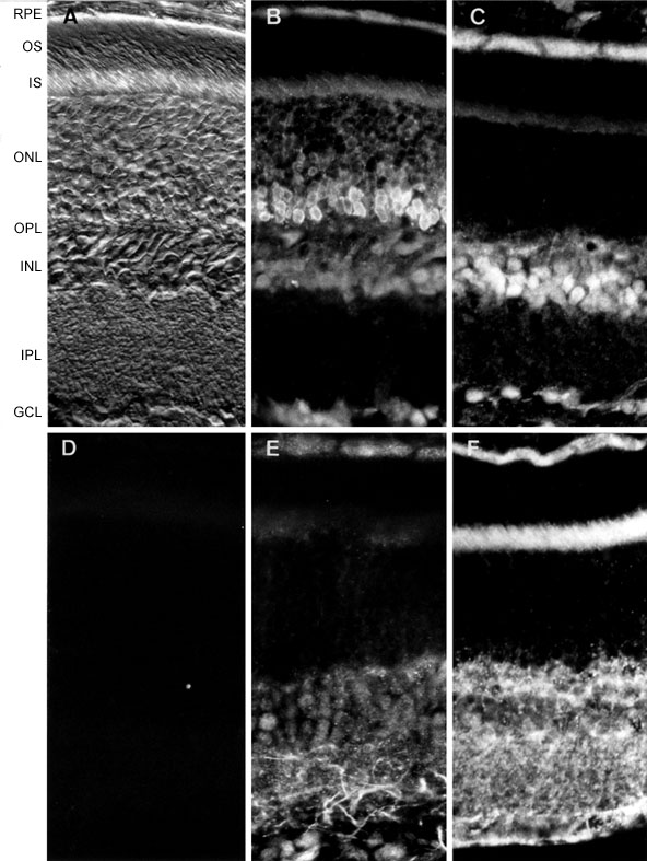
Figure 2. Distribution of FGFRs in adult rat retina ex vivo
Rat retinal sections close to the optic nerve head (shown by differential interference contrast microscopy in A) were stained with antibodies specific for FGFR-1 (B), FGFR-2 (C), FGFR-3 (E), and FGFR-4 (F). While FGFR-1 strongly labeled all nuclear layers, FGFR-2 and -3 showed more intense staining in the inner nuclear layer (INL), inner plexiform layer (IPL) and ganglion cell layer (GCL). FGFR-4 labeling was prominent in the inner retina and also very intense in photoreceptor inner segments (IS). D: Representative control section in which the primary antibody (in this case FGFR-4) was pre-adsorbed with the corresponding immunizing peptide prior to labeling. Additional abbreviations: ONL, outer nuclear layer; OPL, outer plexiform layer; OS, photoreceptor outer segments; RPE, retinal pigmented epithelium. Magnification x280.