![]() Figure 10 of
Kinkl, Mol Vis 2002;
8:149-160.
Figure 10 of
Kinkl, Mol Vis 2002;
8:149-160.
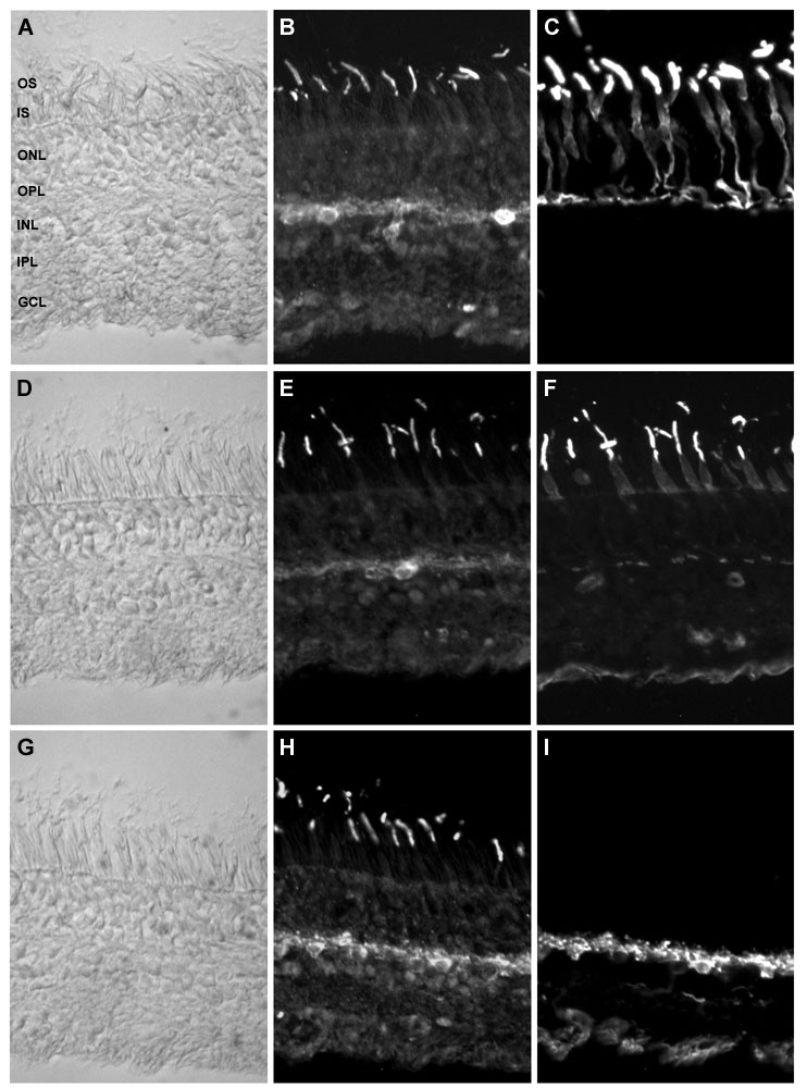
Figure 10. FGFR-2 is abundant in cone outer segments and horizontal cells of human retina ex vivo
A-F: Double immunolabeling of human retinal sections with antibodies specific for FGFR-2 (B, E), 7G6 (C), and PNA (F). FGFR-2 positive cone photoreceptor OS were also labeled by primate cone antibody 7G6 or PNA, showing that FGFR-2 staining was specific for cone OS. G-I: Double immunolabeling with antibodies specific for FGFR-2 (H) and parvalbumin (I). FGFR-2 staining along the margin of the OPL co-localized with parvalbumin-positive horizontal cells. A, D, and G: Differential interference contrast micrographs showing the morphological architecture of the post-mortem human retina. Abbreviations used: GCL, ganglion cell layer; INL, inner nuclear layer; IPL, inner plexiform layer; IS, photoreceptor inner segments; ONL, outer nuclear layer; OPL, outer plexiform layer; OS, photoreceptor outer segments. Magnification x200.