![]() Figure 1 of
Kinkl, Mol Vis 2002;
8:149-160.
Figure 1 of
Kinkl, Mol Vis 2002;
8:149-160.
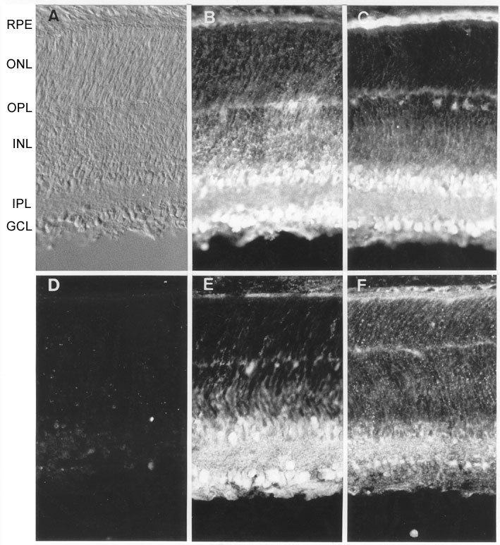
Figure 1. Distribution of FGFRs in PN5 rat retina ex vivo
Rat retinal sections close to the optic nerve head (shown by differential interference contrast microscopy in A) were stained with antibodies specific for FGFR-1 (B), FGFR-2 (C), FGFR-3 (E), and FGFR-4 (F). All four receptors exhibited increased immunoreactivity in the inner retinal layers (INL, inner nuclear layer; IPL, inner plexiform layer; and GCL, ganglion cell layer) compared to the outer nuclear layer (ONL), although FGFR-1 and -4 also revealed moderate staining of this latter. FGFR-1, -2, and -3 showed the most intense labeling within the GCL and vitreal margin of the INL. D: Representative control section in which the primary antibody (in this case FGFR-3) was pre-adsorbed with the corresponding immunizing peptide prior to labeling. Other abbreviations: RPE, retinal pigmented epithelium; OPL, outer plexiform layer. Magnification x280.