![]() Figure 1 of
Rex, Mol Vis 2002;
8:114-118.
Figure 1 of
Rex, Mol Vis 2002;
8:114-118.
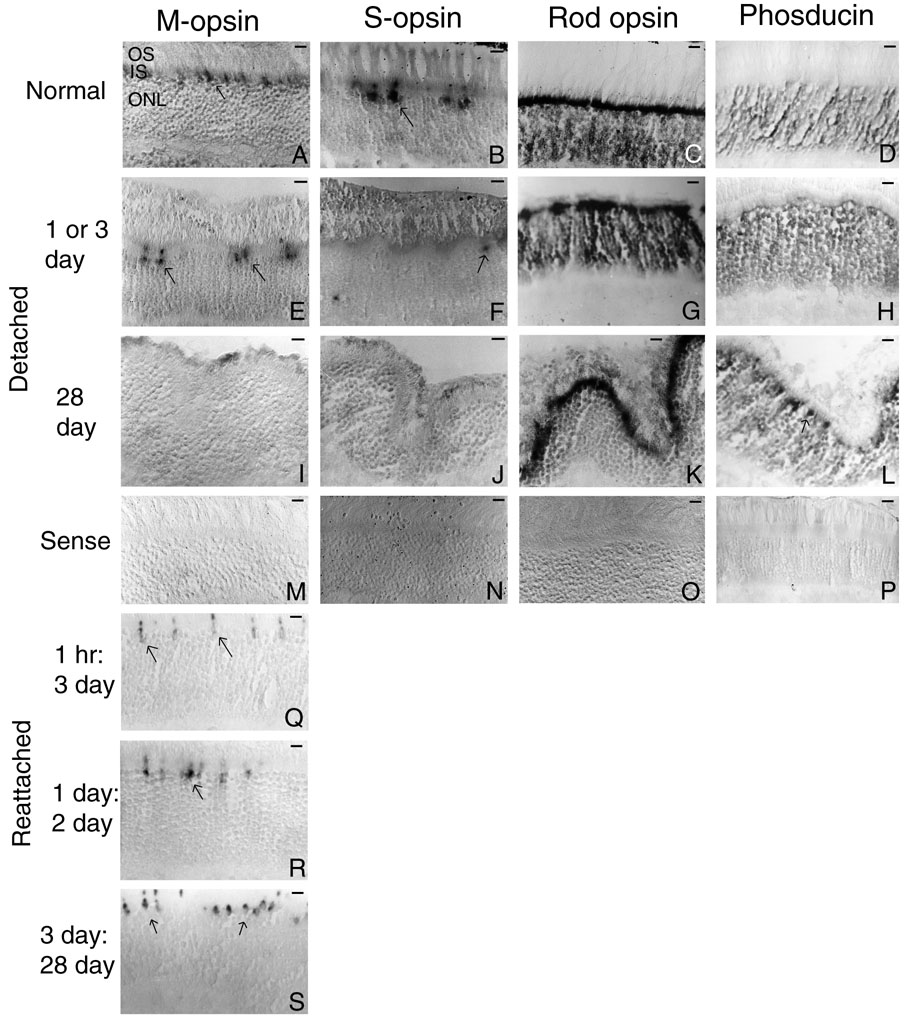
Figure 1. In situ hybridization of normal, detached, and reattached retinas
Light micrographs of normal, detached, and reattached retinas labeled with sense (M, N, O, P) or anti-sense (A-L, Q-S) riboprobe for M-cone opsin (A, E, I, M, Q, R, S), S-cone opsin (B, F, J, N), rod opsin (C, G, K, O), or phosducin (D, H, L, P). Micrographs of 3 day detached retinas (E, F) show a decrease in the number of labeled cones compared to normal (A, B). At 28 days of detachment, no labeled cells were detected (I, J). Micrographs of 1 day detached retinas labeled with either anti-sense rod opsin (G) or phosducin (H) show the labeling levels were similar to that found in normal retina (C, D). Even at 28 days of detachment, the cells remained labeled with these riboprobes (K, L). Anti-sense phosducin darkly labeled cone photoreceptors at 28 days of detachment (L; see arrow). The normal retina sections labeled with sense riboprobe had no background (M-P). After retinal reattachment, the number of cells labeled with M-opsin riboprobe increased to normal (Q-S). Arrows indicate labeled cells. Scale bars represent 22 mm.