![]() Figure 6 of
Wang, Mol Vis 2002;
8:94-101.
Figure 6 of
Wang, Mol Vis 2002;
8:94-101.
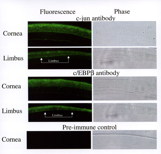
Figure 6. Immunohistochemical staining of mouse corneas
Paraffin sections of central cornea or the limbal region were incubated with rabbit anti-c-jun/AP1, anti-c/EBPβ, and pre-immune control IgG, followed by goat anti-rabbit Alexa Fluor 488. Immunoreactivity was detected using a Nikon eclipse E800 microscope with an epi-fluorescence attachment.