![]() Figure 5 of
Ko, Mol Vis 2002;
8:1-9.
Figure 5 of
Ko, Mol Vis 2002;
8:1-9.
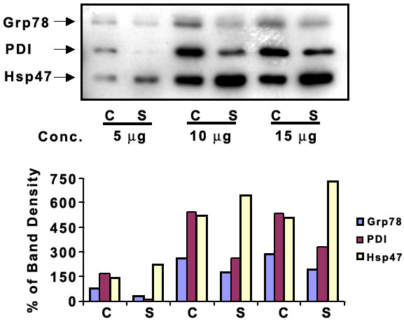
Figure 5. Immunoblot analysis of Grp78/Bip, PDI and Hsp47
Cell lysates prepared from CEC and CSF were separated on a 10% SDS-polyacrylamide gel and transferred to nitrocellulose membrane followed by immunoblotting with mouse anti-PDI, goat anti-Grp78 or mouse anti-Hsp47 antibodies. The relative density of the polypeptide bands was estimated using a one-dimensional image analyzer. Abbreviations: C, CEC; S, CSF; Conc., serial concentration of cell lysates. The data represent two independent experiments.