![]() Figure 3 of
Ko, Mol Vis 2002;
8:1-9.
Figure 3 of
Ko, Mol Vis 2002;
8:1-9.
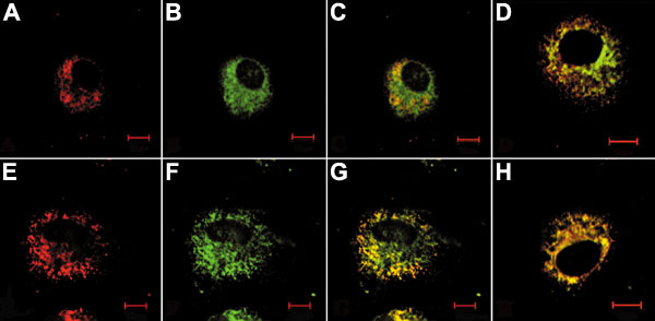
Figure 3. Subcellular localization of procollagen I and type IV collagen with Grp78/BiP
CEC cells were fixed, permeabilized, and stained with antibodies as described in the methods. Some cells were treated with 0.3 mM a,a'-dipyridyl for 2 h prior to fixing and staining (D and H). A: Stained for Grp78/Bip alone. B: Stained for procollagen I alone. C: Merged image of A (red) and B (green). D: Merged image of Grp78/Bip (red) and procollagen I (green) in a,a'-dipyridyl treated cells. E: Stained for Grp78/Bip alone. F: Stained for type IV collagen alone. G: Merged image of E (red) and F (green). H: Merged image of Grp78/Bip (red) and type IV collagen (green) in a,a'-dipyridyl treated cells. Bar represents 10 mm. The data represent four independent experiments.