![]() Figure 1 of
Ko, Mol Vis 2002;
8:1-9.
Figure 1 of
Ko, Mol Vis 2002;
8:1-9.
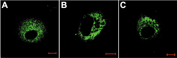
Figure 1. Subcellular localization of P4Ha, PDI and Grp78/Bip
CEC cells were fixed, permeabilized, and stained with antibodies as described in the methods. A: Cells stained for P4Ha; B: Cells stained for PDI; C: Cells stained for Grp78/BiP. Bar represents 10 mm. The data represent three independent experiments.