![]() Figure 4 of
Wagner, Mol Vis 2001;
7:57-62.
Figure 4 of
Wagner, Mol Vis 2001;
7:57-62.
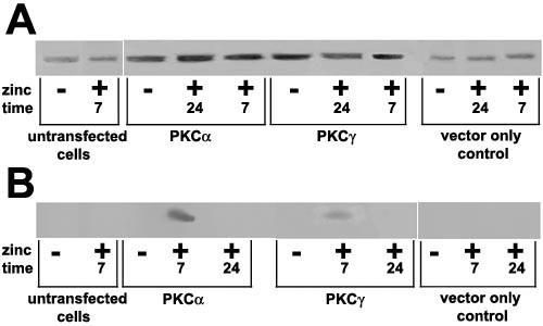
Figure 4. Western blot analysis of aB- and aA-crystallin
Protein (50 mg) from total cell extract was separated by SDS-PAGE and blotted onto a nitrocellulose membrane. The blot was probed with either antisera against aB-crystallin (A) at 1:500 (specific to amino acid residues 117-191) or aA-crystallin (B) at 1:500 (specific to amino acid residues 49-86). aB-crystallin which is normally expressed in the N/N 1003A cell line was detected in all the samples. aA-crystallin which is not expressed in the N/N 1003A cell line was not detected in the untransfected cell line, the cells transfected with empty vector, and cells overexpressing PKCa or PKCg for 24 h. However, aA-crystallin was detected in cells overexpressing PKCa and a lower expression level was observed in cells overexpressing PKCg for 7 days. In the "time" row, the number 7 refers to 7 days of overexpression and the number 24 refers to 24 h of overexpression.