![]() Figure 7 of
Yu, Mol Vis 2001;
7:48-56.
Figure 7 of
Yu, Mol Vis 2001;
7:48-56.
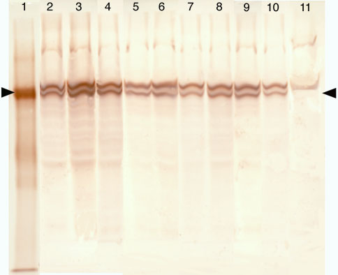
Figure 7. Western blot analysis of fl-Hsc/Hsp70 in normal and light-damaged retinas
Lane 1 is 20 ng of fl-Hsc/Hsp70 from the same preparation used for the intravitreal injections and the arrowhead indicates the prominent immunoreactive band at 70 kD, with very little fluorescein immunoreactivity at other molecular weights. The succeeding lanes are samples of light-damaged and normal retinas collected at increasing intervals after intravitreal injections of fl-Hsc/Hps70, as follows: 2 and 3, replicates of light damaged retinas one h post-injection; 4, normal retina one h post-injection; 5 and 6, light-damaged retinas six h post-injection; 7, normal retina six h post-injection; 8 and 9, light-damaged retinas 24 h post-injection; 10, normal retina 24 h post-injection; 11, normal retina from uninjected eye to show the nonspecific stained bands. The right arrowhead indicates the position of intact fl-Hsc/Hsp70 in the retinal samples, which lined up with the sample of purified fl-Hsc/Hsp70 shown in lane 1. The amount of each retinal sample loaded on the gel was 2.5% of the total retinal homogenate and each eye had received an injection of 10 mg fl-Hsc/Hsp70.