![]() Figure 3 of
Jomary, Mol Vis 2001;
7:36-41.
Figure 3 of
Jomary, Mol Vis 2001;
7:36-41.
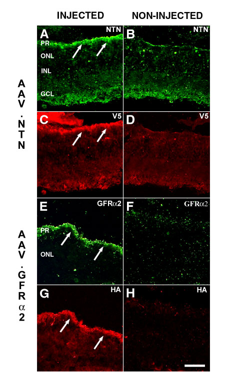
Figure 3. Immunofluorescence analysis of rd mice intraocularly injected with rAAV constructs
Only one eye per animal was injected (panels A, C, E, and G), the other serving as untreated control (panels B, D, F, and H). Total (including endogenous) gene expression is shown by green fluorescence, transgene only by red fluorescence. For eyes injected with AAV.NTN (panels A and C) and their controls (panels B and D), transgene expression was detected using a polyclonal goat anti-human NTN antibody (panels A and B) in conjunction with a monoclonal anti-V5 antibody (panels C and D). In AAV.GFRa-2 injected eyes (panels E and G) and their controls (panels F and H), transgene expression was detected using a polyclonal goat anti-human GFRa-2 antibody (panels E and F) in conjunction with a monoclonal anti-HA antibody (panels G and H). Arrows show co-localized expression of the transgenes and either NTN or GFRa-2 proteins. ONL: outer nuclear layer; INL: inner nuclear layer; GCL: ganglion cell layer; PR: photoreceptors. Bar, 100 mm.