![]() Figure 1 of
Jablonski, Mol Vis 2001;
7:27-35.
Figure 1 of
Jablonski, Mol Vis 2001;
7:27-35.
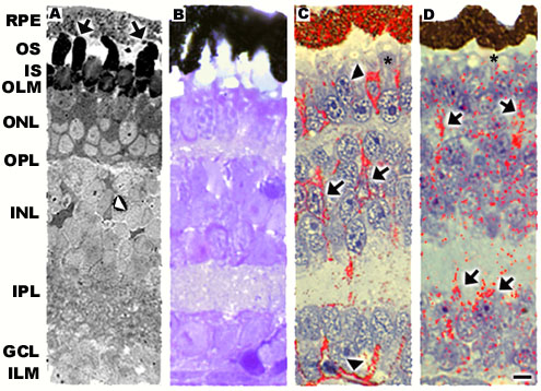
Figure 1. Morphology and immunolocalization patterns of Müller cell-specific proteins
Morphology and immunolocalization patterns of Müller cell-specific proteins of healthy Xenopus laevis retinas with an adherent RPE. In panel A, the tissue was fixed in glutaraldehyde and osmium tetroxide so that the morphology of the tissue including the outer segments is well-preserved. In panels B-D, the tissues were fixed in Davidson fixative that does not stain the membranous components of the photoreceptor outer segment. A: In the presence of the RPE, photoreceptor outer segments were composed of an orderly array of stacked discs (arrows). Some diamond-shaped nuclear profiles were evident in the INL and are likely to be Müller cell nuclei (arrowhead). B: GFAP immunolabeling was not evident in these retinas. C: CRALBP immunolabeled heavily the RPE and Müller cell radial processes. Diamond-shaped immunolabeled profiles were evident in the INL (arrows). Müller cells were heavily labeled at the level of the outer and inner limiting membranes (arrowheads). A low level of CRALBP immunolabeling was present immediately beyond the outer limiting membrane between adjacent IS (asterisk). D: Glutamine synthetase was present throughout Müller cell radial processes with heavy immunopositive labeling in both plexiform layers (arrows). Glutamine synthetase labeling also was apparent immediately beyond the outer limiting membrane (asterisk). RPE = retinal pigment epithelium, OS = outer segment, IS = inner segment, OLM = outer limiting membrane, ONL = outer nuclear layer, OPL = outer plexiform layer, INL = inner nuclear layer, IPL = inner plexiform layer, GCL = ganglion cell layer, ILM = inner limiting membrane. Bar = 10 mm.