![]() Figure 8 of
Chen, Mol Vis 2001;
7:305-313.
Figure 8 of
Chen, Mol Vis 2001;
7:305-313.
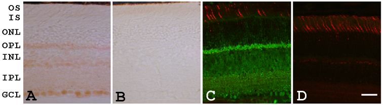
Figure 8. GRK7 is not present in mouse cone outer segments
Localization of GRK7 using UU45APC (A and C, used at 1:1,000 dilution) and pre-absorbed UU45APC (B and D, used at 1:500 dilution) in overnight fixed mouse retina by chromogenic detection (A and B) and by fluorescent detection (C and D). GRK7 is present in the OPL (outer plexiform layer), INL (inner nuclear layer), IPL (inner plexiform layer) and GCL (ganglion cell layer), but absent in the ONL (outer nuclear layer), the IS (inner segments) and the OS (outer segments). Fluorescent double labeling using rhodamine conjugated peanut agglutinin to bind to mouse cone sheaths reveals the absence of GRK7 signal in mouse cone outer segments. Bar represents 30 mm.