![]() Figure 4 of
Riley, Mol Vis 2001;
7:297-304.
Figure 4 of
Riley, Mol Vis 2001;
7:297-304.
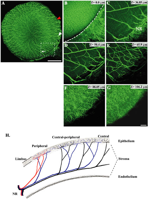
Figure 4. Analysis of nerves at different corneal depths
Confocal images of a whole mount E16 quail cornea immunostained with the TuJ1 monoclonal antibody. A: Whole cornea was scanned at an optimum focal depth to show the positioning of both short (red arrowhead) and long nerves (white arrow and arrowhead). B-G are enlargements of the boxed region in A z-sectioned at different focal depths starting at the basal surface of the endothelium (B), going distally towards the epithelial surface (G) of the cornea. H: Schematic drawing of a cornea cross section showing the distribution of both short nerves (red) and long nerves (blue and black). Dotted line beneath the epithelium denotes interface with Bowman's layer. Dashed line above the endothelium denotes interface with Descemet's layer. The dotted line in (B) demarcates the interface between the cornea and the limbus. Abbreviations: NR, nerve ring. Bars represents 500 mm.