![]() Figure 3 of
Zhao, Mol Vis 2001;
7:277-282.
Figure 3 of
Zhao, Mol Vis 2001;
7:277-282.
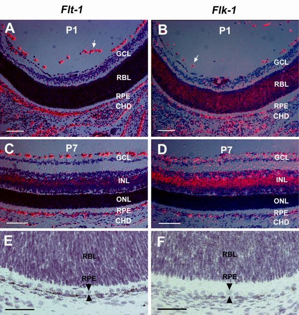
Figure 3. Expression of VEGF receptors in the developing eye
Messenger RNA transcripts of Flt-1 (A, C, E) and Flk-1 (B, D, F) were assayed by in situ hybridizations using 35S-labeled probes. At P1 (A, B), Flt-1 and Flk-1 mRNAs were expressed in the endothelial cells of hyaloid blood vessels (arrows) and in the periocular mesenchyme. Flk-1 mRNA was also expressed in the retinoblast layer (B). At P7 (C, D), Flt-1 and Flk-1 mRNAs were expressed in the vascular endothelial cells at the vitreous surface and in the periocular mesenchyme. The inner nuclear layer of the neural retina expressed low levels of Flt-1 mRNA (C) but high levels of Flk-1 mRNA (D). Bright-field images of P1 eye sections at higher magnification show heavy labeling of silver grains in a subset of the mesenchymal cells adjacent to the RPE expressing Flt-1 (E) and Flk-1 transcripts (F). The RPE layer is marked by a pair of arrowheads (E, F). Abbreviations: GCL, ganglion cell layer; INL, inner nuclear layer; ONL, outer nuclear layer; RBL, retinoblast layer. Scale bars: 100 mm (A, B, C, D) and 50 mm (E, F).