![]() Figure 1 of
Zhao, Mol Vis 2001;
7:277-282.
Figure 1 of
Zhao, Mol Vis 2001;
7:277-282.
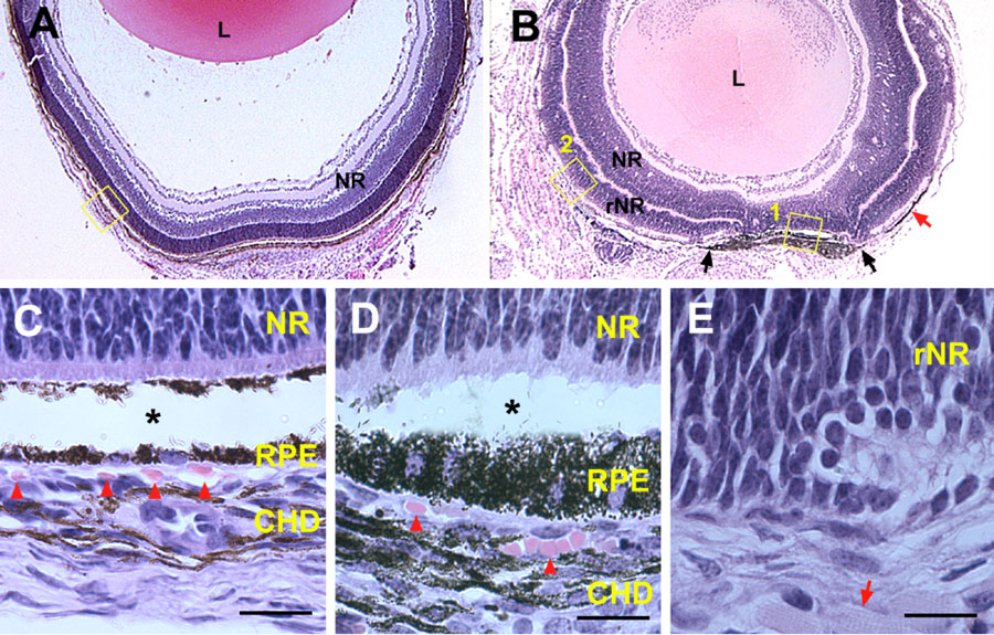
Figure 1. Failure in choroid formation in the absence of the RPE
Hematoxylin-eosin staining of P7 eye sections from wild type (A, C) and TRP2-FGF9 transgenic (B, D, E) mice. In wild type mice, the neural retina (NR) had differentiated and formed three distinct layers (A). In the TRP2-FGF9 transgenic eye (B), the vitreous did not form and the majority of the presumptive RPE was converted to a second layer of neural retina except for a patch of RPE in the posterior region (between the two black arrows in B). The developing choroid adjacent to this region had become pigmented. Thin monolayers of melanocytes were occasionally found in regions where the RPE was absent (red arrow in B). However, no tissues resembling developing choroid were observed in these regions. The boxed region in A is shown in C while boxes 1 and 2 in B are shown in D and E, respectively. In wild type P7 eyes (C), melanocytes already appeared in the developing choroid and small blood vessels and blood cells were present (arrowheads in C). In the transgenic eyes, melanocytes were present in the developing choroid next to the unconverted RPE patch (B, D). Blood vessels were seen in this choroidal tissue (arrowheads in D). In the region where the RPE was absent (box 2 in B), no blood vessels were observed in the presumptive choroid tissue adjacent to the RPE-derived neural retina (rNR) (E). The arrow in E indicates a striated muscle cell. Detachment of neural retina from RPE (asterisks in C, D) was due to artifacts in tissue processing. Abbreviations: CHD, developing choroid; L, lens. Scale bars: 20 mm.