![]() Figure 1 of
Yin, Mol Vis 2001;
7:271-276.
Figure 1 of
Yin, Mol Vis 2001;
7:271-276.
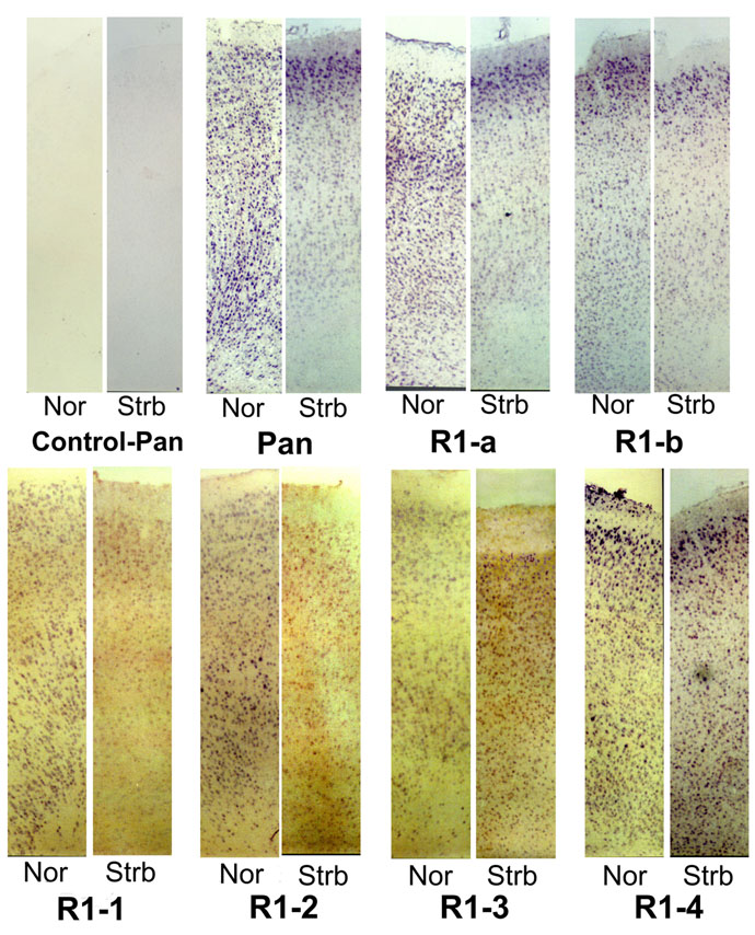
Figure 1. ISH staining of NMDAR1 mRNA isoforms
Light micrographs of the full cortical thickness showing ISH staining for the alternatively spliced isoforms of the NMDAR1 mRNA normal and strabismic cats. There was no signal found by hybridization with the control probe. As expected, the "pan" probe, which detects all the NMDAR1 mRNA species, showed abundant expression in normal visual cortex, but considerably less in strabismic visual cortex. A different distribution of cells stained positively for the heterogeneous variants. The laminar distribution of positive immunostaining for all the hybridized isoforms was most prominent in layer II-III. Fewer positively stained cells were apparent in the strabismic samples except for the deep laminae for the isoform R1-3 probe.