![]() Figure 5 of
Zhou, Mol Vis 2001;
7:253-260.
Figure 5 of
Zhou, Mol Vis 2001;
7:253-260.
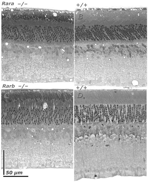
Figure 5. Retinal histology in Rara and Rarb lines
Retinal histology in Rara (upper panels: A, B) and Rarb (lower panels: C, D) lines of mice. Cellular and plexiform layers of retinas of Rara and Rarb knockout mice have apparently normal structure (left panels: A,C). Inner and outer nuclear layers and the plexiform layers are of the appropriate thickness. Subtle differences in thickness of layers and staining quality of the micrographs are caused primarily by section angle. Differences of outer segment "staining" is actually a polarization artifact associated with differential interference contrast optics.