![]() Figure 2 of
Saleh, Mol Vis 2001;
7:240-246.
Figure 2 of
Saleh, Mol Vis 2001;
7:240-246.
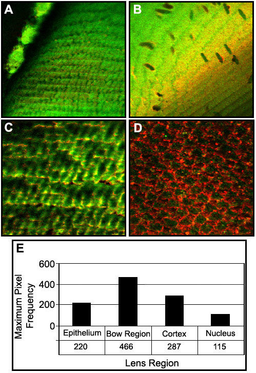
Figure 2. PKC-g and Cx46 in different regions in the lens
Co-localization of PKC-g and Cx46 in different regions in the lens. Lens sections (100 mm thick) were labeled with anti-PKC-g (green) and anti-Cx46 antibodies (red), fixed, then examined with the confocal microscope. Co-localization is demonstrated by a yellow color (overlap of green and red colors). The objective lens was 63x 1.4 oil immersion objective. A: Lens epithelial cells show a lack of PKC-g and Cx46 co-localization. B: Fiber cells of the bow region show PKC-g and Cx46 co-localization. C: Cortical fibers show PKC-g and Cx46 co-localization. D: Nuclear fibers show a lack of PKC-g and Cx46 co-localization. E: Quantitative analysis of pixel intensity of co-localization of PKC-g and Cx46. Background is equal to 100-200 (1% of total pixels). Antisera for PKC-g was used at 1:1000 (Transduction Labs). Cx46 antisera was used at 1:1000 (a gift of Dr. L. Takemoto, Kansas State University).