![]() Figure 1 of
Saleh, Mol Vis 2001;
7:240-246.
Figure 1 of
Saleh, Mol Vis 2001;
7:240-246.
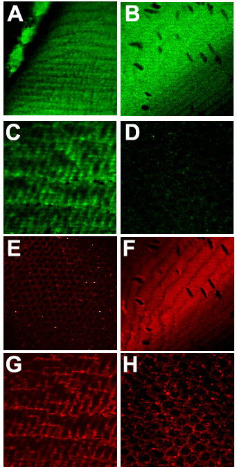
Figure 1. Localization of Cx46 and PKC-g
Individual localization of PKC-g (A-D, green) and Cx46 (E-H, red) in the epithelial layer (A and E), bow region (B and F), cortex (C and G), and nucleus (D and H) of the lens. The objective lens was a 63x 1.4 oil immersion objective. D (PKC-g) and E (Cx46) are similar to background labeling observed using non-immune sera (D is negative for PKC-g, E is negative for Cx46).