![]() Figure 3 of
Burgio, Mol Vis 2001;
7:228-233.
Figure 3 of
Burgio, Mol Vis 2001;
7:228-233.
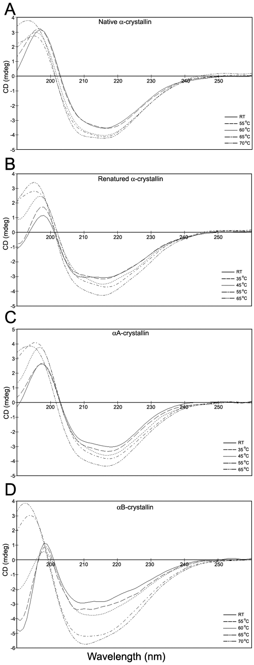
Figure 3. Far-UV CD spectra
Far-UV CD spectra (178-260 nm) were obtained of a-crystallin samples incubated for 1 h at the temperature indicated and allowed to cool to room temperature (RT) before data was collected. All spectra were taken in 0.1 mm cylindrical cells at protein concentrations of 1-1.5 mg/ml and subsequently adjusted to 1 mg/ml. A: Native a-crystallin; B: Renatured a-crystallin; C: aA-Crystallin; D: aB-Crystallin. Samples incubated at temperatures from 35 °C to 50 °C in the native a- and aB-crystallin samples showed no apparent change from the RT spectra and are not shown in A and D. In B and C, only five of the nine spectra taken are presented. The unpresented spectra follow the existing trends and are not included to improve the clarity of the figures.