![]() Figure 2 of
Burgio, Mol Vis 2001;
7:228-233.
Figure 2 of
Burgio, Mol Vis 2001;
7:228-233.
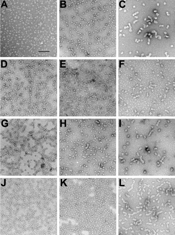
Figure 2. Transmission electron micrographs
Transmission electron micrographs were obtained of a-crystallin samples incubated for 1 h at various temperatures and allowed to cool to room temperature (RT) before being placed on the grid. They were then negatively stained with uranyl acetate and photographed at 50,000x magnification. The solid bar in A represents 100 nm for all of the micrographs. A: Native a-crystallin at room temperature (RT); B: Native a-crystallin at 50 °C; C: Native a-cryatallin at 65 °C; D: Renatured a-crystallin at RT; E: Renatured a-crystallin at 50 °C; F: Renatured a-crystallin at 65 °C; G: aA-Crystallin at room temperature (RT); H: aA-Crystallin at 50 °C; I: aA-Crystallin at 65 °C; J: aB-Crystallin at RT; K: aB-Crystallin at 50 °C; L: aB-Crystallin at 65 °C.