![]() Figure 4 of
Shim, Mol Vis 2001;
7:216-221.
Figure 4 of
Shim, Mol Vis 2001;
7:216-221.
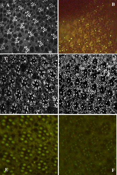
Figure 4. Fluorescence micrographs with PLC antibody
Fluorescence micrographs prepared with PLC antibody, repletion manipulations, and selected mutants. A: 3 days carrot juice replacement of deprived flies, showing the recovery of rhabdomere size, confocal micrograph. B: negative control, the mutant white-eyed norpAP24, standard diet, standard fluorescence micrograph, no rhabdomere fluorescence detected as expected. C: the white-eyed mutant ninaEoI17, standard diet, confocal micrograph; note normal level of PLC detected in R7 rhabdomeres. D: the white-eyed mutant ninaEora, standard diet, confocal micrograph; note normal level of PLC detected in R7. E: the transformant TI-6 rescued from white-eyed norpAP24 by norpA minigene driven by ninaE promoter, showing PLC detected in only R1-6 rhabdomeres, standard diet, standard fluorescence micrograph. F: the transformant TI-6 rescued from white-eyed norpAP24 by norpA minigene driven by ninaE promoter, showing no fluorescence emission, retinoid deprived diet, standard fluorescence micrograph.