![]() Figure 3 of
Shim, Mol Vis 2001;
7:216-221.
Figure 3 of
Shim, Mol Vis 2001;
7:216-221.
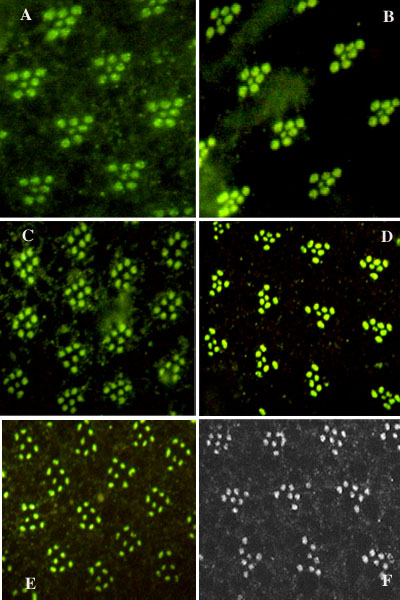
Figure 3. Immunocytochemistry for PLC
Immunocytochemistry for PLC in white-eyed, otherwise wild-type flies, retinoid replete, and retinoid or chromophore deprived. A: standard diet, bright fluorescence in R1-7 rhabdomeres, standard fluorescence micrograph. B: rearing on Sang's medium supplemented with b-carotene from egg to adult (one generation) showing the same bright fluorescence as for normal diet as expected, standard fluorescence micrograph. C: retinoid deprived; note that rhabdomeres fluoresce brightly but that the size of rhabdomeres may be decreased, standard fluorescence micrograph. D: rearing on yeast-glucose diet, showing same features as for deprived diet, standard fluorescence micrograph. E: rearing on Sang's medium supplemented with Beef Brain-Heart Infusion from egg to adult (one generation) showing the same features as for retinoid deprived diet, standard fluorescence micrograph. F: rearing with retinoic acid supplemented Sang's medium for one generation showing same features as for deprived diet, confocal micrograph.