![]() Figure 1 of
Hu, Mol Vis 2001;
7:14-19.
Figure 1 of
Hu, Mol Vis 2001;
7:14-19.
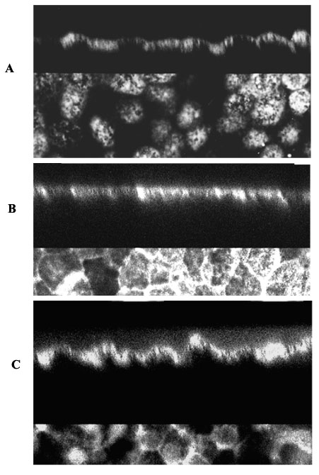
Figure 1. Expression of Na,K ATPase by human RPE cultured in three different media
RPE cells were grown on polycarbonate filters for 2 months. The upper portion of each image is a cross-section of the RPE monolayer using the Phi-Z mode of the confocal microscope. The lower portion of each image shows a single optical section taken in the Z axis. A: CEM replacement medium, the optical section is at the apical surface. B: DMEM/F12 culture medium. The optical section is taken near the basal surface. C: DMEM with high glucose. The optical section is taken at a point intermediate between the apical and basal surface.