![]() Figure 2 of
Zhu, Mol Vis 2001;
7:210-215.
Figure 2 of
Zhu, Mol Vis 2001;
7:210-215.
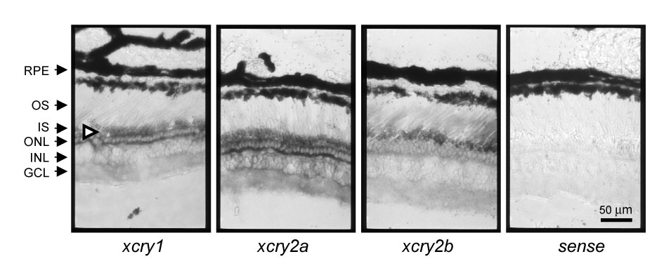
Figure 2. Spatial analysis of xcry mRNA expression in the Xenopus retina
Xenopus eyes were dissected and fixed at ZT 2 and 14 mm cryosections were prepared. In situ hybridization analysis was done with digoxygenin-labeled antisense (left three) and sense (right) xcry probes. (Only the sense of xcry1 was shown here. Similar results were obtained for both xcry2a and xcry2b sense probes.) The white arrowhead emphasizes the heavy in situ labeling in the inner segments of the photoreceptor cells. Retinal cell layers are labeled on the left: RPE, retinal pigment epithelium; OS, photoreceptor outer segments; IS, photoreceptor inner segment; ONL, outer nuclear layer (photoreceptor cell nuclei); INL, inner nuclear layer; and GCL, ganglion layer.