![]() Figure 6 of
Morgans, Mol Vis 2001;
7:202-209.
Figure 6 of
Morgans, Mol Vis 2001;
7:202-209.
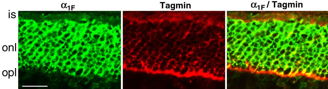
Figure 6. Double labeling for anti-a1F(rat) and synaptotagmin.
Double labeling for a1F and a synaptic vesicle marker in the outer retina demonstrates that a1F is restricted to the photoreceptor cell bodies and is excluded from the synaptic terminals. Rat retina sections were doubled labeled for rat a1F shown in green, and synaptotagmin (Tagmin) shown in red. The images were superimposed (right-most panel) to reveal areas of co-localization, which appear yellow. Abbreviations: IS, inner segments of photoreceptors; ONL, outer nuclear layer; OPL, outer plexiform layer.