![]() Figure 5 of
Morgans, Mol Vis 2001;
7:202-209.
Figure 5 of
Morgans, Mol Vis 2001;
7:202-209.
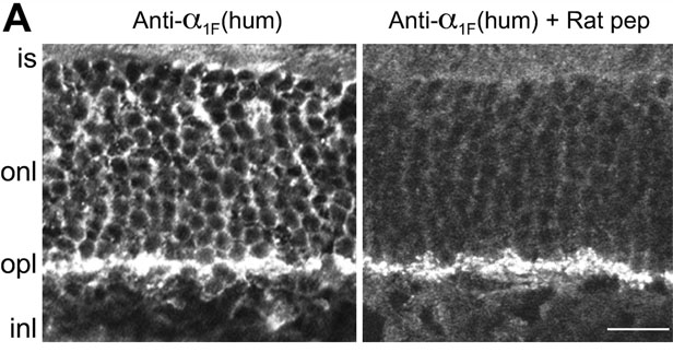
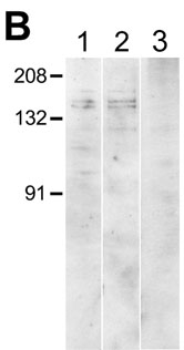
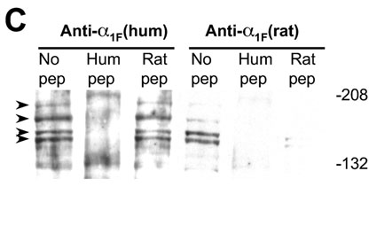
Figure 5. Evidence for additional a1F isoforms or a1F-like calcium channel subunits in the rat retina.
A: Anti-a1F(human) strongly labels puncta in the OPL in addition to labeling the ONL (left panel). Preincubation of anti-a1F(human) with the rat peptide blocks staining of the ONL, but has little effect on the OPL staining (right panel). Size bar is 12 mm. Abbreviations are as follows: is, inner segments; onl, outer nuclear layer; opl, outer plexiform layer; inl, inner nuclear layer. B: On western blots of rat retinal membrane proteins, both anti-a1F(human), shown in lane 1, and anti-a1F(rat), shown in lane 2, label predominant bands at 140 and 150 kD. Lane 3 shows a blot of brain membrane proteins probed with anti-a1F(human); no bands are detected. Positions of molecular weight standards are indicated on the left. C: Western blots of whole retina proteins with both anti-a1F(rat) and anti-a1F(human) reveal, in addition to the bands at 140 and 150 kD, two higher molecular weight bands at 170 and 190 kD (lanes marked No pep, bands indicated with arrowheads). Preincubation of either anti-a1F(rat) or anti-a1F(human) with the humana1F peptide (Hum pep) completely eliminated all four bands for both antibody fractions. Preincubation of anti-a1F(rat) with an equivalent concentration of the rat a1F peptide (Rat pep) also blocked labeling of the four bands. In contrast, all four bands persisted following preincubation of anti-a1F(human) with the rat a1F peptide. Positions of molecular weight standards are indicated on the right.