![]() Figure 4 of
Morgans, Mol Vis 2001;
7:202-209.
Figure 4 of
Morgans, Mol Vis 2001;
7:202-209.
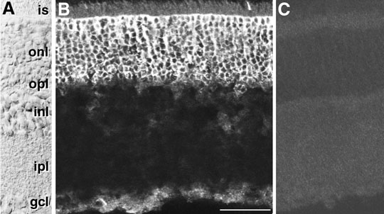
Figure 4. Localisation of anti-a1F(rat) in the rat retina.
Staining with anti-a1F(rat) shows that the a1F calcium channel is localized to the ONL in the rat retina (B). The staining is completely blocked by pre-incubation of the antibody with the rat a1F peptide demonstrating the specificity of the antibody (C). A Nomarski image of a rat retina section with the layers labeled is shown in panel A. Abbreviations: IS, inner segments of photoreceptors; ONL, outer nuclear layer; OPL, outer plexiform layer; INL, inner nuclear layer; IPL, inner plexiform layer; GCL, ganglion cell layer. The 40 mm size bar in panel B applies to all panels.