![]() Figure 7 of
Schlamp, Mol Vis 2001;
7:192-201.
Figure 7 of
Schlamp, Mol Vis 2001;
7:192-201.
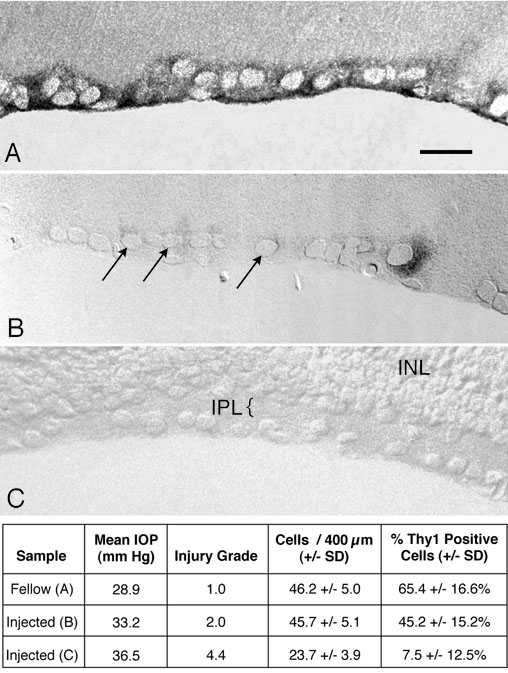
Figure 7. Localization of Thy1 expression in hypertensive eyes
In situ hybridization of Thy1 in a normal rat eye and two eyes with ocular hypertension. A table with specific data for each of the three specimens shown is below the micrographs. A: High magnification Nomarski image of the central superior retina of a control rat eye. Typically, this region contains approximately 46 cells/400 mm of retina of which approximately 65% exhibit robust levels of Thy1 mRNA. B: Similar region of an eye exposed to a mean IOP of 33.2 mm Hg for 35 days. This eye exhibited moderate damage to the optic nerve (approximately 10% fiber loss), but no significant loss of cells in the retina relative to control eyes (p > 0.2, Mann Whitney test). The number of Thy1-positive cells is significantly reduced (p < 0.01) and the pattern of staining shows both strongly positive and weakly positive cells (arrows) all of which were scored in the quantification of these specimens. C: Thy1 staining in a retina from an eye with a mean IOP of 36.5 mm Hg and severe optic nerve damage. The retina had sustained notable damage including the loss of approximately 50% of the cells in the ganglion cell layer and marked thinning of the inner plexiform layer (IPL). Few Thy1-positive cells were evident among the cells remaining in the ganglion cell layer. INL, inner nuclear layer. Scale bar, 20 mm.