![]() Figure 2 of
Schlamp, Mol Vis 2001;
7:192-201.
Figure 2 of
Schlamp, Mol Vis 2001;
7:192-201.
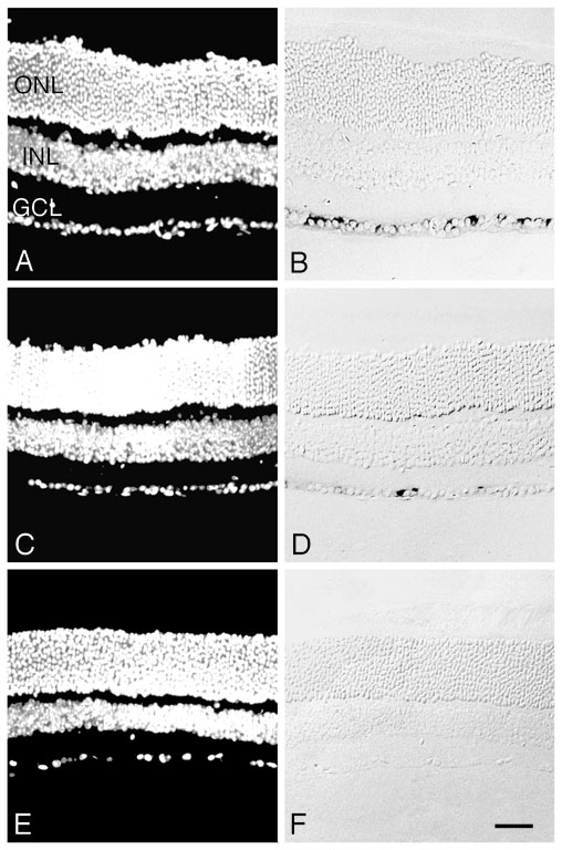
Figure 2. Localization of Thy1 expression after optic nerve crush
In situ hybridization showing the loss of Thy1 mRNA in retinal ganglion cells after optic nerve crush. DAPI-stained and Nomarski images of the same sections of central retina are shown in the left and right panels, respectively. The sections shown are of 14 day control retina (A, B) and 7 days (C, D), and 14 days (E, F) after crush. The majority of cells in the ganglion cell layer (GCL) are positive for Thy1 in control retinas and up to 3 days after surgery (data not shown), but only a few positive cells can be detected by 7 days. No positive cells are detected by this procedure by 14 days. ONL, outer nuclear layer; INL, inner nuclear layer. Scale bar, 20 mm.