![]() Figure 6 of
Sulik, Mol Vis 2001;
7:184-191.
Figure 6 of
Sulik, Mol Vis 2001;
7:184-191.
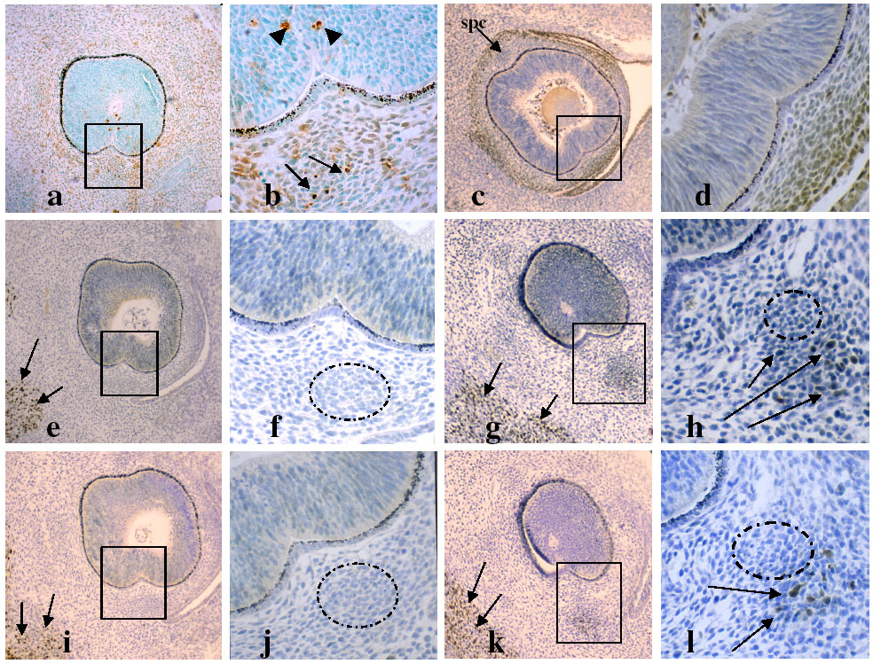
Figure 6. Muscle-specific and AP-2 labeling on gestational day 12.5
A,B: At this time in development, some TUNEL-positive cells remain present in the periocular condensations. The boxed area includes the ipc which is shown at high magnification in B with arrows indicating TUNEL-positive ipc cells and arrowheads indicating dead cells in the choroid fissure. C,D: AP-2 labeling surrounds the eye, but is less intense in the superior condensation (arrow in C) than in the others or the remainder of the scleral primordia (boxed area in C is shown at higher magnification in D). E-H: Myo D labeling of histological sections is positive in developing facial muscles (arrows in E,G) at a plane just behind the lens (E,F) and at a deeper plane (G,H) in the extraocular muscles (arrows in H). (Boxed areas in E,G are shown at higher magnification in F,H, respectively). Cells of the ipc are circled in F,H and are negative for MyoD. I-L: Myogenin labeling in the facial muscles (arrows in I,K) and extraocular muscles (arrows in L) is comparable to that of MyoD. The cells of the ipc are circled in J,L and are negative for muscle protein labeling. The ipc is in contact, at its outer margins, with the positively-labeled extraocular muscle precursors (H,L).