![]() Figure 5 of
Sulik, Mol Vis 2001;
7:184-191.
Figure 5 of
Sulik, Mol Vis 2001;
7:184-191.
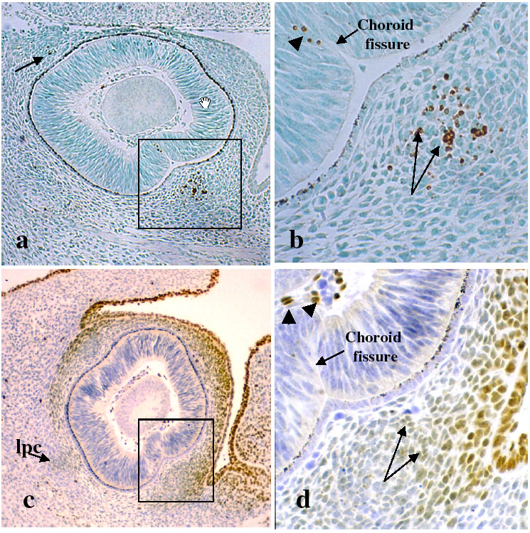
Figure 5. TUNEL and AP-2 labeling of gestational day 12 embryos
A: The ipc, which is shown in the boxed area and at higher magnification in B, is intensely TUNEL positive. Labeled cells are also evident in the spc (arrow in A) and choroid fissure (arrowhead in B). C: AP-2 labeling surrounds and includes all of the peripheral condensations except the lpc. The boxed area in C is shown at higher magnification in D. AP-2 labeling is less intense in the cells of the condensations (arrows in D) than in the scleral precursors, surface ectoderm, and vitreous (arrowheads in D.)