![]() Figure 2 of
Sulik, Mol Vis 2001;
7:184-191.
Figure 2 of
Sulik, Mol Vis 2001;
7:184-191.
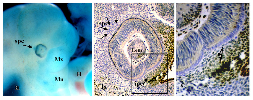
Figure 2. Apoptosis and AP-2 staining in gestational day 11.5 mouse embryos
A: NBS uptake can be seen in the position of the spc indicating programmed cell death in the day 11.5 mouse embryos. B: AP-2 labeling is evident in the surface ectoderm, and portions of the periocular mesenchyme including the medial and inferior peripheral condensations. Boxed area in (B) is shown at higher magnification in panel C. C: AP-2-labeled cells in the ipc are indicated by arrows. AP-2 labeling is also apparent in the lens and vitreous, while precursors of the choroid, immediately adjacent to the optic cup are unlabeled. (An artifactual tear is present in the region of the ipc.) H = heart, Mx = maxillary prominence of first branchial arch, Mn = mandibular prominence of first branchial arch.