![]() Figure 1 of
Sulik, Mol Vis 2001;
7:184-191.
Figure 1 of
Sulik, Mol Vis 2001;
7:184-191.
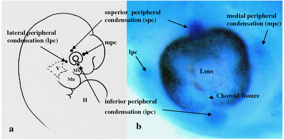
Figure 1. Mesenchymal condensations at the periphery of the developing eye
A: The positions of these peripheral periocular condensations are illustrated in a line drawing of an embryo head (modified from Gilbert [2]). B: They are also shown in a gestational day 12 NBS-stained mouse embryo whose mother had been acutely exposed to ethanol. Ethanol exposure accentuates stain uptake/apoptotic cell death and aids in visualizing the position of the condensations. V = trigeminal ganglion, H = heart, Mx = maxillary prominence of first branchial arch, Mn = mandibular prominence of first branchial arch.