![]() Figure 1 of
Karakousis, Mol Vis 2001;
7:154-163.
Figure 1 of
Karakousis, Mol Vis 2001;
7:154-163.
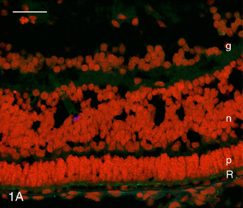
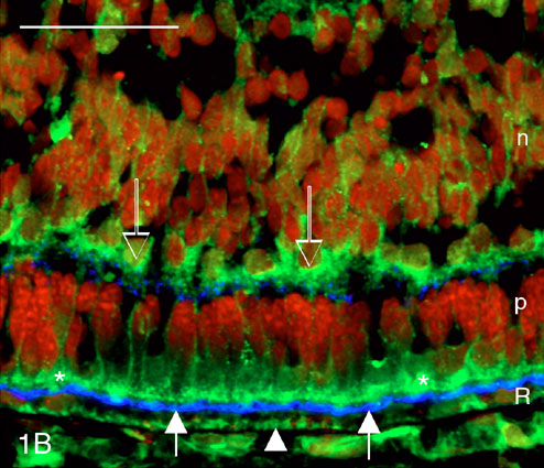
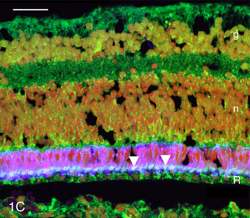
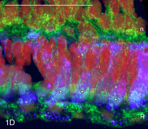
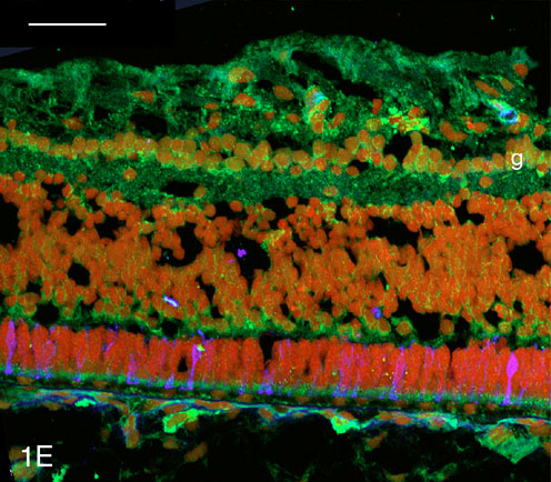
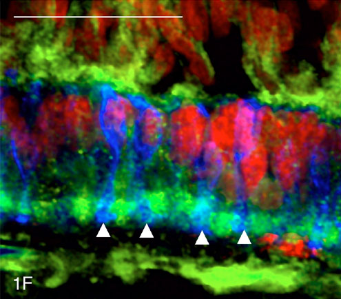
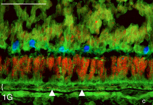
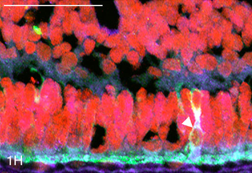
Figure 1. Immunocytochemical labeling of central part of developing human retina at age 21.5 Fwks
A: Control section treated with no primary antibody but with Cy-2- and Cy-5-labeled secondary antibodies shows lack of autofluorescence of the retina and RPE (R). p, differentiating photoreceptors; n, inner nuclear layer; g, ganglion cell layer. B: Double labeling with pAb anti-PEDF (green) and mAb anti-IRBP (blue). IRBP is restricted to a narrow band (arrow) of interphotoreceptor matrix between the photoreceptors (p) and retinal pigment epithelium (R). Note PEDF-positive inner segments (*) of the differentiating photoreceptors. PEDF labeling is also found in cytoplasmic granules in the RPE (arrowhead) and in cells (open arrows) in the outer part of the inner nuclear layer (n). C: Retina treated with the cone-specific mAb 7G6 (blue) and pAb anti-PEDF (green). Note cone inner segments (arrowheads) double labeled (cyan) with 7G6 and anti-PEDF. Many ganglion cells (g) are also PEDF-positive, as are scattered cells in the inner nuclear layer (n), especially the outer part. D: Higher magnification of C. Note cone inner segments (*) double labeled (cyan) with pAb anti-PEDF and mAb 7G6. PEDF-positive cells are present in the outer part of the inner nuclear layer (n). E: Developing rods are positive for rhodopsin (blue) but negative with pAb anti-PEDF (green). Note PEDF-positive cells in the ganglion cell (g) layer. F: Higher magnification of rods (arrowheads) that are positive with mAb anti-rhodopsin (blue) but negative with pAb anti-PEDF (green). G: Immunolabeling with pAb anti-PEDF (green) of fine granules (arrowheads) in the RPE (bracket). PEDF-labeling is also found in choroidal cells (c), but not in the developing horizontal cells, which have been labeled (blue) with anti-calbindin. H: Monoclonal anti-PEDF (blue) labels the interphotoreceptor matrix, which is also positive with pAb anti-IRBP (green) producing a cyan color. The cytoplasm of one cone (arrowhead) is positive with both anti-IRBP and anti-PEDF. Cell nuclei are stained (red) with propidium iodide. R, retinal pigment epithelium; p, photoreceptor layer; n, inner nuclear layer; g, ganglion cell layer. Bars = 40 mm.