![]() Figure 6 of
Wagner, Mol Vis 2001;
7:138-144.
Figure 6 of
Wagner, Mol Vis 2001;
7:138-144.
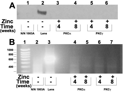
Figure 6. Analysis to detect g-crystallin in lentoid bodies
g-Crystallin was not detected in normal N/N 1003A lens epithelial cells (A, lane 1; B, lane 2). Western blot analysis (A) and RT-PCR (B) did not detect significant expression of g-crystallin after the overexpression of either PKC isoform. In B, lane 1 is a molecular size marker and lane 3 is mouse lens cDNA for g-crystallin.