![]() Figure 5 of
Wagner, Mol Vis 2001;
7:138-144.
Figure 5 of
Wagner, Mol Vis 2001;
7:138-144.
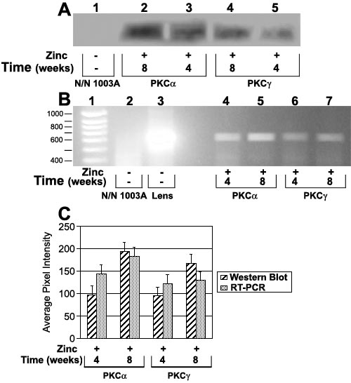
Figure 5. Analysis to detect b-crystallin in lentoid bodies
Normal cultures of N/N 1003A lens epithelial cells did not express b-crystallin by Western blot analysis (A, lane 1) or by RT-PCR (B, lane 2). b-Crystallin was detected in slightly lower levels (statistically significant) in 4 week old lentoid bodies compared to 8 week old lentoid bodies (A, lane 2 vs 3 and lane 4 vs 5; B, lane 4 vs 5 and lane 6 vs 7), and shown by densitometric analysis of the blots and RT-PCR (C). Lentoid bodies formed after PKCa overexpression expressed b-crystallin at significantly higher levels (statistically significant) than those formed after PKCg overexpression at 8 weeks (A, lane 2 vs 4; B, lane 5 vs 7), and as shown by densitometric analysis of the blots and RT-PCR (C; a=0.05). In B, lane 1 is a molecular size marker and lane 3 is mouse lens cDNA for b-crystallin.