![]() Figure 3 of
Wagner, Mol Vis 2001;
7:138-144.
Figure 3 of
Wagner, Mol Vis 2001;
7:138-144.
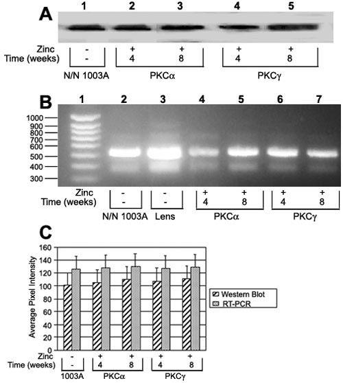
Figure 3. Analysis to detect aB-crystallin in lentoid bodies
Western blot analysis (A) and RT-PCR (B) detected aB-crystallin in normal cultures of N/N 1003A lens epithelial cells (A, lane 1; B, lane 2) and in 4 and 8 week old lentoid bodies caused by the overexpression of PKCa (A, lanes 2 and 3; B, lanes 4 and 5) and of PKCg (A, lanes 4 and 5; B, lanes 6 and 7). The amount of expression was not significantly different (a=0.05) for both time periods nor were there any significant differences in aB-crystallin between the cells overexpressing either PKCa or PKCg shown by densitometric analysis (C). In B, lane 1 is a molecular size marker and lane 3 is mouse lens cDNA for aB-crystallin.