![]() Figure 2 of
Wagner, Mol Vis 2001;
7:138-144.
Figure 2 of
Wagner, Mol Vis 2001;
7:138-144.
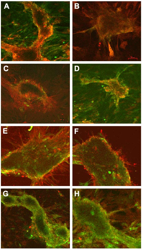
Figure 2. Immunofluorescent staining of lentoid bodies
N/N 1003A lens epithelial cells were plated on 6 well tissue culture plates and PKCa or PKCg overexpression was induced with 20 mM zinc acetate. Lentoid bodies were allowed to form and further differentiate. The lentoid structures were fixed and stained with antibodies to PKCa and PKCg and to aB-, aA-, b-, and g-crystallin. aB-Crystallin was found throughout the differentiation process for the overexpression of PKCa (A) and PKCg (B). Two weeks after lentoid body formation, aA-crystallin was expressed in lentoid bodies overexpressing PKCa (C) and 3 weeks after lentoid structure formation aA-crystallin was expressed in lentoid bodies overexpressing PKCg (D). b-crystallin expression began after 3.5 weeks for PKCa overexpression (E) and 5.5 weeks for PKCg overexpression (F). Lentoid bodies did not express g-crystallin in lentoid bodies formed from the overexpression of PKCa (G) and PKCg (H).