![]() Figure 6 of
Shaw, Mol Vis 2001;
7:6-13.
Figure 6 of
Shaw, Mol Vis 2001;
7:6-13.
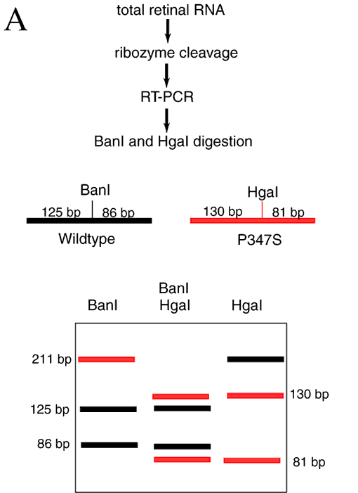
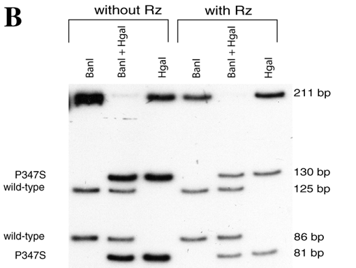
Figure 6. Cleavage of P347S mRNA
A: The protocol and logic of the RT-PCR reaction performed to monitor cleavage of the porcine P347S mRNA by the porcine P347S hammerhead ribozyme. RT-PCR fragments that represent wild type sequences are in black and RT-PCR fragments derived from the P347S mutant mRNA are in red. Complete digestion of the RT-PCR products with BanI leaves a 211 base-pair fragment that represents total P347S mutant mRNA. Complete digestion of the RT-PCR products with HgaI leaves a 211 base-pair fragment that represents wild type mRNA. B: Autoradiograph of a non-denaturing 6% polyacrylamide gel used to separate the BanI and HgaI digested RT-PCR products produced after the P347S hammerhead ribozyme cleavage reaction.

