![]() Figure 3 of
Shaw, Mol Vis 2001;
7:6-13.
Figure 3 of
Shaw, Mol Vis 2001;
7:6-13.
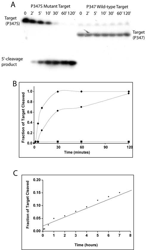
Figure 3. Specificity of the P347S hammerhead ribozyme
A: Autoradiograph of a 10% polyacrylamide 8 M urea gel used to separate the products of the porcine hammerhead ribozyme cleavage of the P347S and P347 target RNA oligonucleotides. B: Graphical representation of the data obtained from the gel in Panel A shows the fraction of the RNA target cleaved over time. Diamonds represent the results obtained from cleavage of the P347S RNA target oligonucleotide and boxes represent the results obtained from cleavage of the P347 RNA wild type target oligonucleotide. The circles represent data obtained from a time course experiment on the cloned version of the P347S target. C: Graphical representation of data obtained from cleavage by the human P347S hammerhead ribozyme on the human P347S RNA target oligonucleotide.
