![]() Figure 1 of
Timmers, Mol Vis 2001;
7:131-137.
Figure 1 of
Timmers, Mol Vis 2001;
7:131-137.
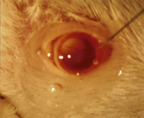
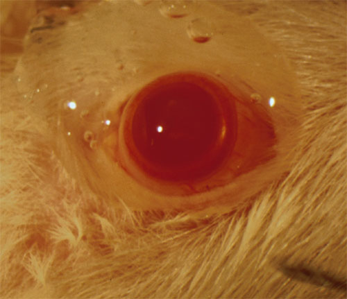
Figure 1. Subretinal injections in rodent with anterior approach
Series of images of Sprague Dawley rat eyes during the injection procedure. The rats were anesthetized with a mixture of ketamine and xylazine; phenylephrine and a topical anesthetic were applied to the eyes.
A: Complete mydriasis prior to injection.
B: Corneal puncture of the eye with a 28 gauge needle. Approximately 70 to 100% of the bevel has been advanced into the anterior chamber, the point of puncture was approximately 1.5 mm medial to the pupillary margin.
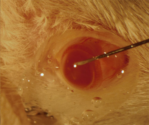
C: The 33 gauge blunt needle tip in the anterior chamber of the punctured eye. Subsequently, the needle was angled to point slightly nasally and guided posteriorly into the eye toward the injection site. Upon penetration of the retina, a PBS-fluorescein mixture was deposited subretinally.
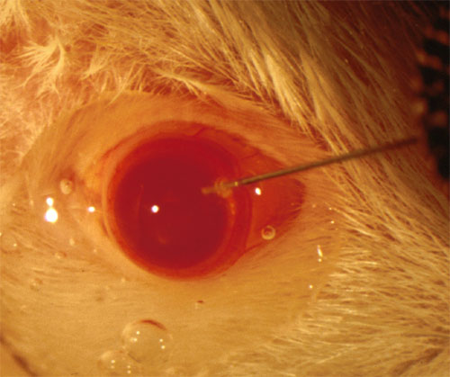
D: After injection of 2 ml of PBS-fluorescein mixture, the retinal bleb is visible with the dye located subretinally. Note the retinal blood vessel overlying the green bleb indicative of a successful subretinal injection.