![]() Figure 7 of
Gilliland, Mol Vis 2001;
7:120-130.
Figure 7 of
Gilliland, Mol Vis 2001;
7:120-130.
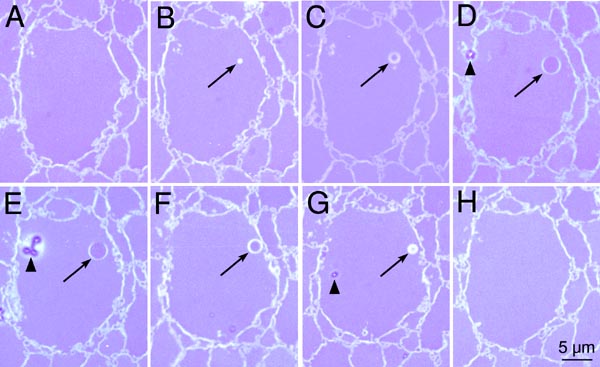
Figure 7. Serial sections of an MLB
Serial sections of an MLB in a cell in the embryonic nucleus. The MLB is not present in A or H. Based on 0.5-mm-thick light microscopy serial sections, the MLB (arrow) increases in size in B through D, achieving its widest diameter (3.4 mm) in E and decreases in size in F and G. The appearance of this typical MLB in serial sections suggests that its overall shape is spherical. The arrowheads indicate sectioning/staining artifacts.